
Objective—Mebendazole and other anti-parasitic drugs are being used off-prescription based on social media and unofficial accounts of their anti-cancer activity. The purpose of this study was to conduct a controlled evaluation of mebendazole’s therapeutic efficacy in cell culture and in vivo models of ovarian cancer.
Epithelial ovarian cancer is the most lethal gynecologic cancer, largely because it has a high predilection for advanced-stage diagnosis (such as Stage 3C) and inevitably develops resistance to chemotherapy despite initially high response rates. While front-line chemotherapy regimens yield an initial remission rate of approximately 80%, fewer than 50% of patients survive past five years.
Current maintenance therapies used post-chemotherapy include bevacizumab (a VEGF-targeted angiogenesis inhibitor) and PARP inhibitors. Although these maintenance strategies prolong progression-free survival, they have a limited or uncertain impact on overall survival while imposing substantial toxicities, side effects, and financial expenses. Consequently, there is an urgent clinical need for novel, well-tolerated, and cost-effective maintenance therapies.
The Molecular Barrier: TP53 Mutations
A major bottleneck in treating ovarian cancer is that the vast majority of cases harbor TP53 null or missense mutations. In epithelial ovarian cancer, missense mutations frequently lead to a “gain-of-function” phenotype that actively drives resistance to chemotherapy-induced apoptosis.
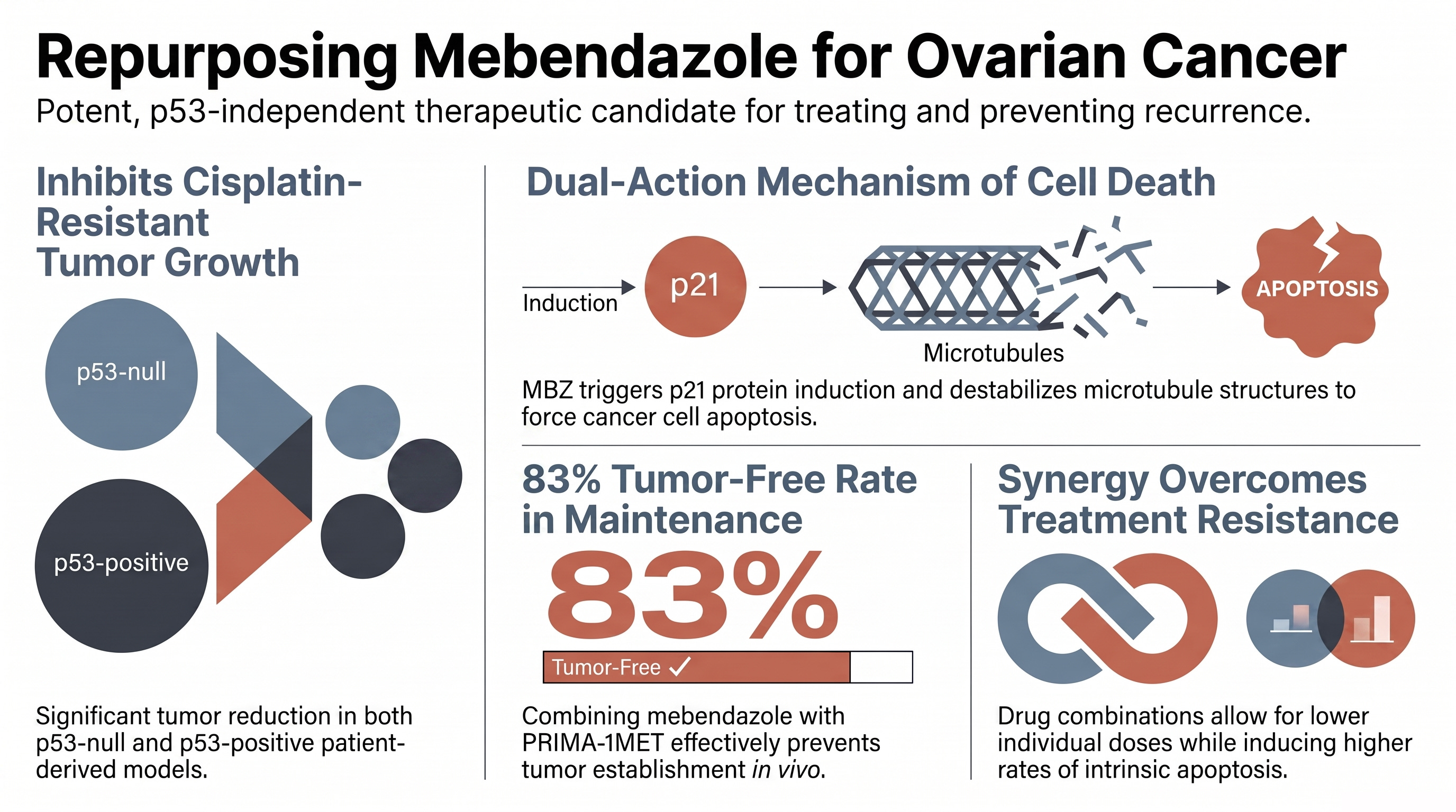
This genetic landscape makes the anti-parasitic drug mebendazole (MBZ) a highly attractive candidate, as its primary anti-tumor mechanisms operate independently of functional p53.
Mebendazole’s Anti-Tumor Mechanisms in Ovarian Cancer
Controlled preclinical evaluations of mebendazole reveal that it is a potent inhibitor of ovarian cancer growth, utilizing a dual-action mechanism:
Tubulin Depolymerization: Mebendazole functions as a microtubule-destabilizing agent. It directly binds to and depolymerizes tubulin, disrupting the cellular microtubule network across various ovarian cancer cell lines.
p53-Independent p21 Induction: Rather than relying on p53 to activate cell cycle arrest, mebendazole significantly upregulates the cyclin-dependent kinase inhibitor p21 regardless of the cell’s p53 mutation status.
Intrinsic Apoptosis Activation: This multi-targeted disruption triggers the cleavages of initiator caspase-9, executioner caspase-3, and PARP-1, forcing aggressive ovarian cancer cells to undergo intrinsic apoptosis.
In vitro, mebendazole inhibits growth at nanomolar concentrations (IC50 values ranging from 400 nM to 1.7 μM) across a broad panel of ovarian cancer lines, including A2780 (p53 wild-type), SKOV3 (p53 null), ES2, MES-OV, and isogenic mutant sublines (SKOV3 R273H and R248W). It also significantly suppresses clonogenic survival and colony formation in these cell lines.
Overcoming Cisplatin Resistance In Vivo
To validate these findings in highly clinically relevant models, researchers evaluated mebendazole in patient-derived xenograft (PDX) models. These models (PDX-0003, which is p53 null, and PDX-0030, which is p53 positive/missense mutant) were derived directly from Stage 3C high-grade serous ovarian cancer patients with recurrent, platinum-resistant disease.
Tumor Inhibition: Oral mebendazole (administered via oral gavage at 10, 25, or 50 mg/kg three times per week for three weeks) significantly inhibited tumor growth in both the p53 null and p53 missense mutant platinum-resistant PDX models.
Dose-Responsive Efficacy: Mixed-effects analysis confirmed that mebendazole’s therapeutic efficacy was highly dose-responsive in both tumor models.
Toxicity Profile: While the highest dose (50 mg/kg) caused a minor, transient reduction in mouse body weight (~5% weight loss, which is well below the 10% maximum tolerated threshold), histological analysis of liver and kidney tissue showed no evidence of systemic organ toxicity.
Preventing Recurrence: Mebendazole as Maintenance Therapy
Because prolonged maintenance therapy requires highly tolerable agents with minimal toxicity, researchers modeled post-chemotherapy maintenance by administering therapies before tumor establishment in an orthotopic intraperitoneal MES-OV (R282W p53 mutant) mouse model.
Standalone Maintenance: Mebendazole monotherapy (50 mg/kg, adjusted to every other day to prevent initial weight loss) successfully prevented tumor establishment, yielding a 67% tumor-free rate compared to 0% in untreated controls.
Synergy with PRIMA-1MET (APR-246): To enhance efficacy against missense mutant p53, mebendazole was combined with the mutant p53 reactivator PRIMA-1MET. In cell culture, this drug combination showed synergistic cytotoxicity and significantly increased intrinsic apoptosis.
Maximizing Tumor-Free Rates: In the in vivo maintenance model, the combination of mebendazole and PRIMA-1MET achieved an outstanding 83% tumor-free rate. Additive logistic regression modeling confirmed that mebendazole was the primary driver associated with preventing tumor establishment.
These robust preclinical results provide a strong scientific rationale for initiating human clinical trials to evaluate mebendazole as a low-cost, low-toxicity maintenance therapy to prolong progression-free survival in ovarian cancer patients after failing platinum-based chemotherapies.
We evaluated the efficacy of combining mebendazole (MBZ) with the mutant p53 reactivator PRIMA-1MET (APR-246) in both in vivo maintenance models and in vitro assays, demonstrating a clear therapeutic advantage over either drug alone.
1. Prevention of Tumor Establishment (In Vivo Maintenance)
In an orthotopic intraperitoneal mouse model designed to simulate post-chemotherapy maintenance, the combination of mebendazole and PRIMA-1MET achieved the highest rate of preventing tumor establishment:
Untreated Control: 0% tumor-free rate.
PRIMA-1MET alone: 25% tumor-free rate.
Mebendazole alone: 67% tumor-free rate.
Combination Treatment: 83% tumor-free rate.
Statistical Nuances:
Pairwise Superiority: Fisher’s exact test confirmed that the combination treatment was statistically more effective at preventing tumor growth than both the untreated control (p=0.015) and PRIMA-1MET alone (p=0.043).
The Main Efficacy Driver: While the combination yielded the highest percentage (83%), an additive logistic regression model showed that mebendazole was the dominant driver of preventing tumor establishment (p=0.005, odds ratio = 27.535).
No Detected Synergy In Vivo: At or under the doses tested in this animal model, the authors did not formally observe a statistically significant additive or synergistic interaction between the two drugs. The authors noted that further testing with varied dose combinations is needed to fully characterize potential in vivo synergy.
2. Synergistic Cytotoxicity (In Vitro)
Unlike the in vivo model, cell culture experiments demonstrated true pharmacological synergy across the board:
Synergistic Interaction: Isobologram analyses yielded Combination Index (CI) values indicating clear synergistic interaction between mebendazole and PRIMA-1MET across all tested ovarian cancer cell lines, regardless of wild-type, missense-mutant, or null p53 profiles.
Dose Reduction Advantage: Dose Reduction Index (DRI) values confirmed that combining the two agents allowed both mebendazole and PRIMA-1MET to be administered at significantly lower doses than would be required as single-drug treatments to achieve the same therapeutic effect.
3. Amplified Programmed Cell Death (Apoptosis)
The combination treatment was significantly more effective at driving cancer cells into programmed cell death:
Greater Apoptotic Response: In cell culture, the combined regimen triggered significantly higher levels of intrinsic apoptosis than either drug alone.
Enhanced Caspase Activation: This was verified by a substantial increase in the cleavage of initiator caspase-9, executioner caspase-3, and the DNA repair enzyme PARP-1.
Source:
Potential and mechanism of mebendazole for treatment and maintenance of ovarian cancer
PMID: 33131904, PMCID: PMC8820236, DOI: 10.1016/j.ygyno.2020.10.010
2021 Jan. doi: 10.1016/j.ygyno.2020.10.010. Epub 2020 Oct 31.