
Introduction
What if an ancient Chinese remedy that revolutionized malaria treatment holds the secret to defeating drug-resistant cancers? Discovered in the leaves of sweet wormwood, artemisinin earned its discoverer a Nobel Prize in 2015—and today, cutting-edge oncology research is revealing its surprising power to selectively hunt down and destroy tumor cells.
In this article, you will explore artemisinin's journey from traditional plant extractions to modern cancer biology. By the end of this piece, you will be able to:
Trace the history and chemical evolution of artemisinin and its semisynthetic derivatives.
Explain the iron-dependent bioactivation mechanism that allows artemisinins to selectively target cancer cells.
Differentiate the primary mechanisms of action underlying artemisinin anticancer toxicity, including ROS generation, apoptosis, cell cycle arrest, and ferroptosis.
Evaluate the clinical evidence supporting artemisinin combination therapies and their chemosensitizing effects in oncology.
The Iron-Triggered Trap: How Artemisinin Fights Cancer
From Ancient Herb to Nobel Prize: The Origins and Chemistry of Artemisinins
Our story begins over two millennia ago in traditional Chinese medicine, where the herb Artemisia annua (known as Qinghao or sweet wormwood) was used as an infusion to treat fevers, jaundice, and hemorrhoids. However, modern scientific research began in 1972 during a secret military initiative in Beijing known as Project 523. Led by pharmaceutical chemist Youyou Tu, researchers isolated the active compound—artemisinin (or Qinghaosu)—from dried Artemisia annua leaves. This groundbreaking discovery earned Tu Youyou the 2015 Nobel Prize in Physiology or Medicine.
Chemically, artemisinin is a sesquiterpene lactone featuring a highly unusual 1,2,4-trioxane ring system. Nestled within this structure is an essential functional group: an endoperoxide bridge (an oxygen–oxygen bond). If this peroxide bridge is removed—as in the synthetic compound deoxyartemisinin—biological activity is almost completely eliminated.
Despite its therapeutic power, natural artemisinin has pharmacokinetic drawbacks: low water/oil solubility, poor oral bioavailability, and a short in vivo half-life (~2.5 hours). To solve these issues, scientists developed several generations of semisynthetic derivatives derived from dihydroartemisinin (DHA), the primary active metabolite of artemisinin:
Artesunate: A water-soluble hemisuccinate derivative widely used in oral, intravenous, and rectal formulations.
Artemether & Arteether: Lipophilic ether derivatives used in oil-based intramuscular injections or oral formulations.
Artemisone: A second-generation amino-artemisinin derivative designed for improved stability, longer half-life, and lower toxicity.
These derivatives are up to five times more potent against both malaria parasites (Plasmodium falciparum) and malignant tumor cells.

Under the Hood: How Artemisinins Selectively Destroy Cancer Cells
How can an antimalarial drug destroy cancer cells without harming healthy tissue? The secret lies in a clever chemical Trojan Horse mechanism involving iron metabolism.
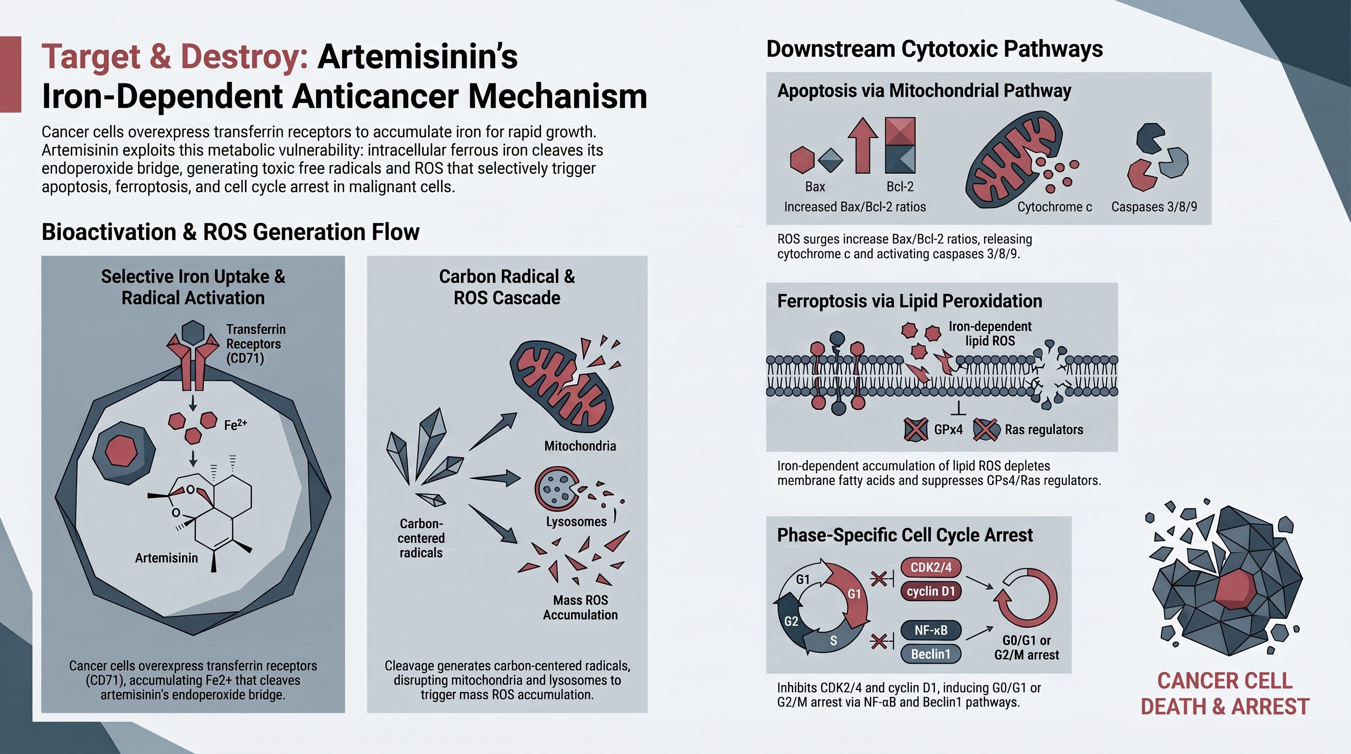
Rapidly multiplying cancer cells need large amounts of iron to synthesize DNA and sustain growth. To capture this iron, tumor cells overexpress Transferrin Receptors (TfR) on their surface. For example, leukemia (CCRF-CEM) cells express TfR on up to 95% of their population, compared with ~1% on normal monocytes. When artemisinin enters a cancer cell's iron-rich intracellular environment, ferrous iron (Fe2+) or heme reacts with the drug’s endoperoxide bridge, breaking the oxygen-oxygen bond and releasing highly potent carbon-centered free radicals and Reactive Oxygen Species (ROS).
Once generated, these toxic ROS molecules unleash multi-targeted destruction within the cancer cell:
Oxidative Stress & Organelle Damage: ROS severely damage cellular membranes, proteins, and DNA. They disrupt lysosomal integrity and trigger mitochondrial dysfunction, releasing cytochrome c into the cytosol.
Induction of Apoptosis: Activated caspases (caspase-3, -8, and -9) drive programmed cell death through the intrinsic pathway by increasing the pro-apoptotic Bax/Bcl-2 ratio.
Ferroptosis: Dihydroartemisinin can trigger a specialized form of iron-dependent, non-apoptotic cell death called ferroptosis. This is marked by lipid ROS accumulation, depletion of membrane polyunsaturated fatty acids, and downregulation of key ferroptotic regulators like GPx4 and Ras.
Cell Cycle Arrest: ARTs alter key regulatory enzymes, trapping cancer cells in the phase (by downregulating Cyclin-Dependent Kinase 4 [CDK4], Cyclin D1, and disrupting signaling) or the phase (by upregulating Beclin1).
Inhibition of Angiogenesis: ARTs starve solid tumors of nutrients by downregulating pro-angiogenic factors such as VEGF (Vascular Endothelial Growth Factor) and CD31, preventing new blood vessel growth.
Power in Partnerships: ARTs in Combination Chemotherapy
Modern oncology relies heavily on combination chemotherapy to maximize tumor destruction while minimizing drug resistance and systemic toxicity. Because artemisinins act through mechanisms distinct from traditional chemotherapeutics, adding them to existing regimens often produces strong synergistic effects and chemosensitization—making previously drug-resistant tumors responsive again.
Key combination studies highlighted in recent research include:
Pancreatic Cancer (DHA + Gemcitabine): Dihydroartemisinin inactivates the transcription factor, which is often induced by gemcitabine and contributes to drug resistance. Combining DHA with gemcitabine increases apoptosis by 2- to 4-fold in pancreatic cancer cells (Panc-1, BxPC-3) and significantly reduces tumor volume in vivo.
Glioma (DHA + Temozolomide): In rat C6 glioma cells, DHA potentiates temozolomide-induced apoptosis and necrosis by 177% via ROS generation.
Lung Cancer (DHA + Cyclophosphamide or Cisplatin): In murine Lewis lung carcinoma and human non-small cell lung cancer (NSCLC) models, combination therapy drastically reduced primary tumor volume and completely eliminated spontaneous pulmonary metastases.
Breast Cancer (Artesunate + Epirubicin): Artesunate sensitizes breast cancer cells to epirubicin in an autophagy cascade-dependent manner.
Hepatoma (DHA/Artemisinin + Gemcitabine): Combinations significantly decreased tumor growth in wild-type () and mutant () human hepatoma models (HepG2, Huh-7, Hep3B)
Translating Bench to Bedside: Clinical Evidence and Safety Profile
While hundreds of preclinical studies demonstrate artemisinins' efficacy, human clinical trials are actively translating these findings into practice.
Key published clinical trials include:
Advanced Non-Small Cell Lung Cancer (NSCLC): Zhang et al. conducted a randomized controlled trial of 120 patients evaluating intravenous artesunate added to standard chemotherapy (vinorelbine + cisplatin). The artesunate group achieved a significantly higher Disease Control Rate (88.2% vs. 72.7%) and a longer median time to disease progression (24 weeks vs. 20 weeks) without added toxicity.
Colorectal Cancer (Preoperative Artesunate): A randomized, double-blind, placebo-controlled trial led by Krishna et al. treated stage II/III CRC patients with oral artesunate (200 mg/day for 14 days) prior to surgery. Artesunate significantly reduced Ki-67 expression (a cellular proliferation marker), reduced circulating CEA tumor marker levels, and achieved an 89% 3-year recurrence-free survival rate compared with 50% in the placebo group.
Advanced Cervical Cancer: Oral artemisinol-R (DHA) given for 28 days induced clinical remission in all 10 enrolled patients, reducing tumor markers (EGFR, Ki-67) and tumor blood vessel density (CD31).
Safety & Tolerability Profile: Artemisinins have a remarkably favorable safety profile, supported by decades of antimalarial use. In oncology trials, daily oral doses up to 200 mg/day (~2.2–3.9 mg/kg/day) are generally well-tolerated. Minor, reversible side effects occasionally reported include transient neutropenia, mild anemia, abdominal discomfort, and vertigo. Routine hematological monitoring is recommended for extended regimens to ensure safe administration.
Summary
Artemisinin and its derivatives (ARTs) are a promising, safe, low-cost class of plant-derived therapeutics that exploit cancer cells’ natural hunger for iron to induce targeted ROS toxicity, cell-cycle arrest, apoptosis, and ferroptosis.
Key Takeaways:
Structure & Origins: Extracted from Artemisia annua (Qinghao), artemisinin contains an essential endoperoxide bridge responsible for its cytotoxic activity. Key semisynthetic derivatives include dihydroartemisinin (DHA), artesunate, and artemether.
Selective Targeting: Cancer cells overexpress Transferrin Receptors (TfR) to import iron, which reacts with artemisinin’s endoperoxide bridge to generate toxic carbon-centered free radicals and ROS.
Multimodal Mechanisms: ARTs kill cancer cells by inducing mitochondrial apoptosis, iron-dependent ferroptosis, cell cycle arrest, and downregulating pro-angiogenic VEGF/CD31 signals.
Chemosensitization: ARTs work synergistically with standard chemotherapy drugs (e.g., gemcitabine, temozolomide, cisplatin), overcoming drug resistance and suppressing metastases.
Clinical Promise: Phase 1 and 2 clinical trials confirm that oral/IV artesunate and DHA are well-tolerated and significantly improve disease control rates and recurrence-free survival in lung, colorectal, breast, and cervical cancers.
Cheng, C., Ho, W. E., Guan, S. P., Kong, L. R., Hafiz, W., Goh, F. Y., Leung, B. P., & Wong, W. S. F. (2011). Anti-malarial drug artesunate attenuates experimental allergic asthma via inhibition of the phosphoinositide 3-kinase/Akt pathway. PLOS ONE, 6(6), Article e20932.
[Source: pone.0020932.pdf]Crespo-Ortiz, M. P., & Wei, M. Q. (2012). Antitumor activity of artemisinin and its derivatives: From a well-known antimalarial agent to a potential anticancer drug. Journal of Biomedicine and Biotechnology, 2012, Article 247597. https://doi.org/10.1155/2012/247597
[Source: BioMed Research International - 2011 - Crespo-Ortiz...pdf]Krishna, S., Bustamante, L., Haynes, R. K., & Staines, H. M. (2008). Artemisinins: Their growing importance in medicine. Trends in Pharmacological Sciences, 29(10), 520–527.
[Source: PIIS0165614708001648.pdf]Krishna, S., Ganapathi, S., Chis Ster, I., Saeed, M. E. M., Cowan, M., Finlayson, C., Kovacsevics, H., Jansen, H., Kremsner, P. G., Efferth, T., & Kumar, D. (2015). A randomized, double blind, placebo-controlled pilot study of oral artesunate therapy for colorectal cancer. EBioMedicine, 2(1), 82–90. https://doi.org/10.1016/j.ebiom.2014.11.010
[Source: PIIS2352396414000346 (1).pdf]Li, Y. (2012). Qinghaosu (artemisinin): Chemistry and pharmacology. Acta Pharmacologica Sinica, 33(9), 1141–1146.
[Source: aps2012104a.pdf]Li, Z., Li, Q., Wu, J., Wang, M., & Yu, J. (2016). Artemisinin and its derivatives as a repurposing anticancer agent: What else do we need to do? Molecules, 21(10), Article 1331.
[Source: molecules-21-01331-v2.pdf]Liu, L., et al. (2015). Antitumor effect of artesunate on human esophageal carcinoma cells in vitro and in vivo. Molecular Medicine Reports, 12(1), 1465–1471.
[Source: mmr_12_1_1465_PDF.pdf]Liu, T., Liu, Q.-L., et al. (2013). Targeted cytotoxicity of artesunate towards retinoblastoma cells via CD71-mediated endocytosis. Oncology Reports, 30(3), 1473–1480.
[Source: or_30_3_1473_PDF.pdf]Slezakova, S., & Ruda-Kucerova, J. (2017). Anticancer activity of artemisinin and its derivatives. Anticancer Research, 37(11), 5995–6003. https://doi.org/10.21873/anticanres.12046
[Source: 5995.full.pdf]