
1. Study Overview and Background Context
Fenbendazole (FBZ) is an established benzimidazole anthelmintic widely utilized in veterinary and human medicine to control parasitic helminth infections. FBZ exerts its antiparasitic effects by binding to β-tubulin, disrupting microtubule equilibrium, and inhibiting tubulin polymerization. Because of its selective microtubule-binding profile, FBZ has increasingly attracted interest as a candidate for drug repurposing in oncology chemotherapy.
Conventional antineoplastic agents that target the microtubule cytoskeleton—such as Vincristine (VC) and Paclitaxel—exert antitumor activity by disrupting mitotic spindle formation and microtubule dynamics. However, these clinical agents frequently cause severe off-target cytotoxicity in healthy tissues and induce profound immunosuppression. Prolonged or repeated cycles of standard chemotherapy impair lymphocyte function and significantly compromise T cell-mediated immune responses. Consequently, rigorous evaluation of FBZ's differential cytotoxicity between malignant neoplastic cells and normal primary immune cells is essential before clinical translation.
This report provides a technical synthesis of experimental data comparing the cytotoxic, metabolic, and functional effects of FBZ with vincristine in a murine T-cell lymphoma cell line (EL-4) and normal primary mouse spleen cells (splenocytes).

2. Experimental Methodology and Assay Specifications
2.1 Cell Lines & Spleen Cell Harvesting
Cell Line Origin: Mouse lymphoma EL-4 cells were obtained from the Korean Cell Line Bank and cultured in RPMI 1640 medium supplemented with 10% fetal bovine serum (FBS), 100 U/mL penicillin-streptomycin, and 2 mM L-glutamine.
Spleen Cell Preparation: Spleens harvested from C57BL/6 and BALB/c mice (7–12 weeks old) were physically disrupted to yield single-cell suspensions. We lysed red blood cells with ammonium chloride potassium (ACK) lysis buffer (Thermo Fisher Scientific). Following hemolysis, the cell suspension was passed through a 70 m cell strainer and resuspended in complete RPMI 1640 medium containing 10% FBS, L-glutamine, and penicillin-streptomycin.
2.2 Quantitative Assays & Flow Cytometry Protocol
Flow cytometric acquisition was conducted using a CytoFLEX LX flow cytometer (Beckman Coulter) and analyzed using CytExpert software.
Cellular Metabolic Activity (MTT Assay): EL-4 cells and spleen cells were seeded in 96-well plates and incubated with FBZ or VC for 3 days. 3-(4,5-dimethylthiazol-2-yl)-2,5-diphenyltetrazolium bromide (MTT) solution was added to a final concentration of and incubated for 4 hours at 37°C in 5%. Formazan crystals were solubilized using a 10% sodium dodecyl sulfate (SDS) solution for 2 hours, and optical density (O.D.) was read at 570 nm on a Multiskan FC microplate reader (Thermo Fisher Scientific).
Apoptosis Analysis: EL-4 and spleen cells were cultured in 24-well plates with FBZ or VC. Cells were stained with Annexin V-fluorescein isothiocyanate (FITC) (Thermo Fisher Scientific), washed with Annexin V-binding buffer, and incubated with Propidium Iodide (PI; Sigma-Aldrich) for 10 minutes at room temperature in the dark.
Mitochondrial Membrane Potential (MMP): Following 48 hours of drug exposure, cells were incubated with Rhodamine 123 (Sigma-Aldrich) for 30 minutes in the dark. Cells were washed twice with fluorescence-activated cell sorting staining solution (FSS) prior to mean fluorescence intensity (MFI) quantification.
Reactive Oxygen Species (ROS) Generation: Following 72 hours of treatment, cell debris was gated out, and cells were washed with pre-warmed PBS and stained with dichlorofluorescein diacetate (DCFDA; Sigma-Aldrich) for 1 hour at 37°C. We washed the stained cells twice with FSS and quantified oxidized DCF fluorescence intensity.
Cell Cycle Analysis: Following treatment with FBZ or VC, cells were washed with PBS and fixed in 70% cold ethanol for 20 minutes at -20°C. Fixed cells were washed to remove fixative and stained with a PI solution at a final concentration in the presence of RNase.
Functional LPS Reactivity: Spleen cells were pretreated with FBZ for 24 hours. Cells were harvested, washed with culture medium, and viable cells were re-seeded and stimulated with Lipopolysaccharide (LPS, purified from Escherichia coli O26:B6; Sigma-Aldrich) for an additional 48 hours. We then measured metabolic activity via MTT assay.
3. Cellular Metabolic Activity and IC50 Comparative Metrics
FBZ induced a robust, concentration-dependent inhibition of metabolic activity in EL-4 lymphoma cells. Statistically significant reductions in EL-4 metabolic activity were observed across all tested concentrations, displaying a sharp decrease at concentrations exceeding (). In contrast, primary spleen cell metabolic activity experienced only minor reductions at select higher concentrations, demonstrating high baseline tolerance relative to malignant cells. Vincristine displayed a comparable selective pattern, inducing a steep decline in EL-4 metabolic activity while sparing primary spleen cells within equivalent working ranges.
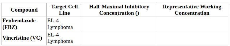
Based on MTT dose-response profiling, the half-maximal inhibitory concentrations () and representative experimental working doses were established for both compounds:
4. Mechanism-Specific Cytotoxicity & Flow Cytometry Findings
Differential Cytotoxicity Summary Profile
4.1 Cell Survival and Apoptosis Induction
Annexin V-FITC / PI flow cytometric analysis revealed marked differences in viability and cell death kinetics between lymphoma cells and primary splenocytes:
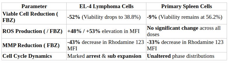
EL-4 Lymphoma Cells: FBZ treatment caused a 52% reduction in viable cells compared with untreated controls. Quadrant analysis showed that FBZ shifted the EL-4 population from 91.5% viable (lower-left) in controls to 38.8% viable, while increasing early apoptotic cells (lower-right) to 43.6% and late apoptotic cells (upper-right) to 17.2% (necrotic cells, upper-left: 0.41%). At FBZ, viable cells shifted to 65.3%, with 27.1% early apoptosis and 6.84% late apoptosis. VC induced a comparable shift: 40.2% viable, 43.5% early apoptotic, 15.7% late apoptotic, and 0.60% necrotic.
Primary Spleen Cells: FBZ treatment caused only a 9% reduction in overall viable cell proportion. Quadrant breakdown for splenocytes exposed to FBZ showed 56.2% viable cells, 39.3% early apoptotic cells, 2.58% late apoptotic cells, and 1.91% necrotic cells (compared to untreated control: 61.9% viable, 35.6% early apoptotic, 1.83% late apoptotic, 0.73% necrotic). At FBZ, splenocyte viability remained high at 43.0% (51.2% early apoptotic, 3.61% late apoptotic, and 2.18% necrotic), confirming that FBZ induces far less catastrophic cell death in primary immune tissue than in lymphoma cells.
4.2 Reactive Oxygen Species (ROS) Production
Intracellular ROS levels were evaluated by measuring oxidized DCFDA mean fluorescence intensity (MFI):
EL-4 Lymphoma Cells: FBZ induced a significant, dose-dependent surge in ROS. Baseline DCFDA MFI in untreated controls was 7,246, increasing slightly to 7,313 and rising sharply to 10,731 (+48%) and 11,101 (+53%). VC induced a comparable elevation in ROS MFI to 9,852.
Primary Spleen Cells: FBZ treatment failed to induce ROS generation in normal splenocytes. Baseline DCFDA MFI in untreated spleen cells was 1,686. Spleen cells treated with FBZ showed MFI values of 1,265, 1,497, and 1,456, respectively, with no statistically significant changes relative to the control. Similarly, VC produced an MFI of 1,522, confirming the total absence of drug-induced oxidative stress in non-malignant spleen tissue.
4.3 Mitochondrial Membrane Potential (MMP) Alteration
MMP loss was evaluated by quantifying Rhodamine 123 MFI:
EL-4 Lymphoma Cells: Baseline Rhodamine 123 MFI in untreated EL-4 cells was 24,434. Exposure to FBZ progressively decreased MMP intensity to 16,021, 15,773, and 13,841 (a 43% reduction relative to control). VC produced a similar MMP reduction, yielding an MFI of 14,912 (a 39% reduction).
Primary Spleen Cells: Baseline Rhodamine 123 MFI in untreated primary spleen cells was 5,537. FBZ exposure yielded MFI values of 4,154, 4,288, and 3,728 (a 33% reduction). VC reduced splenocyte MMP intensity to 3,277 (a 41% reduction).
4.4 Cell Cycle Phase Distribution
Flow cytometric DNA content analysis via PI staining demonstrated that FBZ selectively disrupts cell cycle progression in EL-4 cells while leaving primary spleen cell dynamics intact.
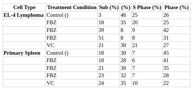
EL-4 Lymphoma Dynamics: FBZ exposure induced pronounced phase arrest (elevating the fraction from 26% to 42%) and reduced the S-phase fraction from 25% to 9%. At FBZ, apoptotic DNA fragmentation predominated, expanding the subpopulation to 51% (compared to 3% in control). VC similarly expanded the subfraction to 21%.
Primary Spleen Dynamics: Spleen cell cycle distributions remained stable across all FBZ concentrations. The subfraction changed minimally from 18% in controls to 21% at and 23% at FBZ, confirming that FBZ does not disrupt cell cycle progression or trigger catastrophic DNA fragmentation in non-malignant splenocytes.
5. Immunological Functionality: Spleen Cell LPS Reactivity
To evaluate whether exposure to FBZ compromises the functional activation capacity of surviving primary immune cells, splenocytes were evaluated using a two-stage pretreatment and mitogenic stimulation protocol:
FBZ Pretreatment: We cultured primary spleen cells with FBZ for 24 hours. Although total viable splenocyte counts decreased by 21% after FBZ pretreatment relative to untreated controls, we harvested sufficient viable cells for functional testing.
Mitogenic LPS Stimulation: Viable splenocytes isolated from pretreatment conditions were re-seeded at equal densities and stimulated with bacterial LPS for 48 hours.
Functional Outcome: Splenocytes pretreated with FBZ across all concentrations displayed robust metabolic activation and proliferative responses following LPS challenge ( relative to non-LPS-treated parallel controls; at ). These results confirm that while FBZ exerts mild cytotoxic effects on overall splenocyte numbers, surviving primary immune cells retain complete immunological reactivity and functional responsiveness to pathogenic/mitogenic stimuli.
6. Summary Synthesis: FBZ vs. VC Cytotoxicity Profile
Antineoplastic Efficacy on Lymphoma (EL-4):
FBZ exhibits potent, targeted antineoplastic activity against EL-4 T-cell lymphoma.
FBZ shares key mechanistic pathways with Vincristine: tubulin binding, induction of intracellular ROS (+53%), loss of mitochondrial membrane potential (-43%), prominent cell cycle arrest (42% at ), and apoptotic subexpansion (51% at ).
Spleen Immune Cell Toxicity:
FBZ exhibits striking differential cytotoxicity, showing high tolerability in normal primary spleen cells compared to lymphoma cells ( viability reduction in splenocytes vs. in EL-4 cells at ).
Unlike in malignant lymphoma cells, FBZ produces no significant ROS elevation and induces no cell cycle arrest or substantial subaccumulation in primary spleen tissue.
Preservation of Immune Function:
FBZ exposure preserves functional immunocompetence in healthy primary lymphocytes. Viable splenocytes pretreated with FBZ maintain full mitogenic activation and metabolic response capabilities upon bacterial LPS stimulation.
These findings indicate that Fenbendazole offers a favorable therapeutic index, achieving anticancer potency comparable to conventional tubulin-binding chemotherapeutics like Vincristine while minimizing off-target immunosuppressive toxicities.