
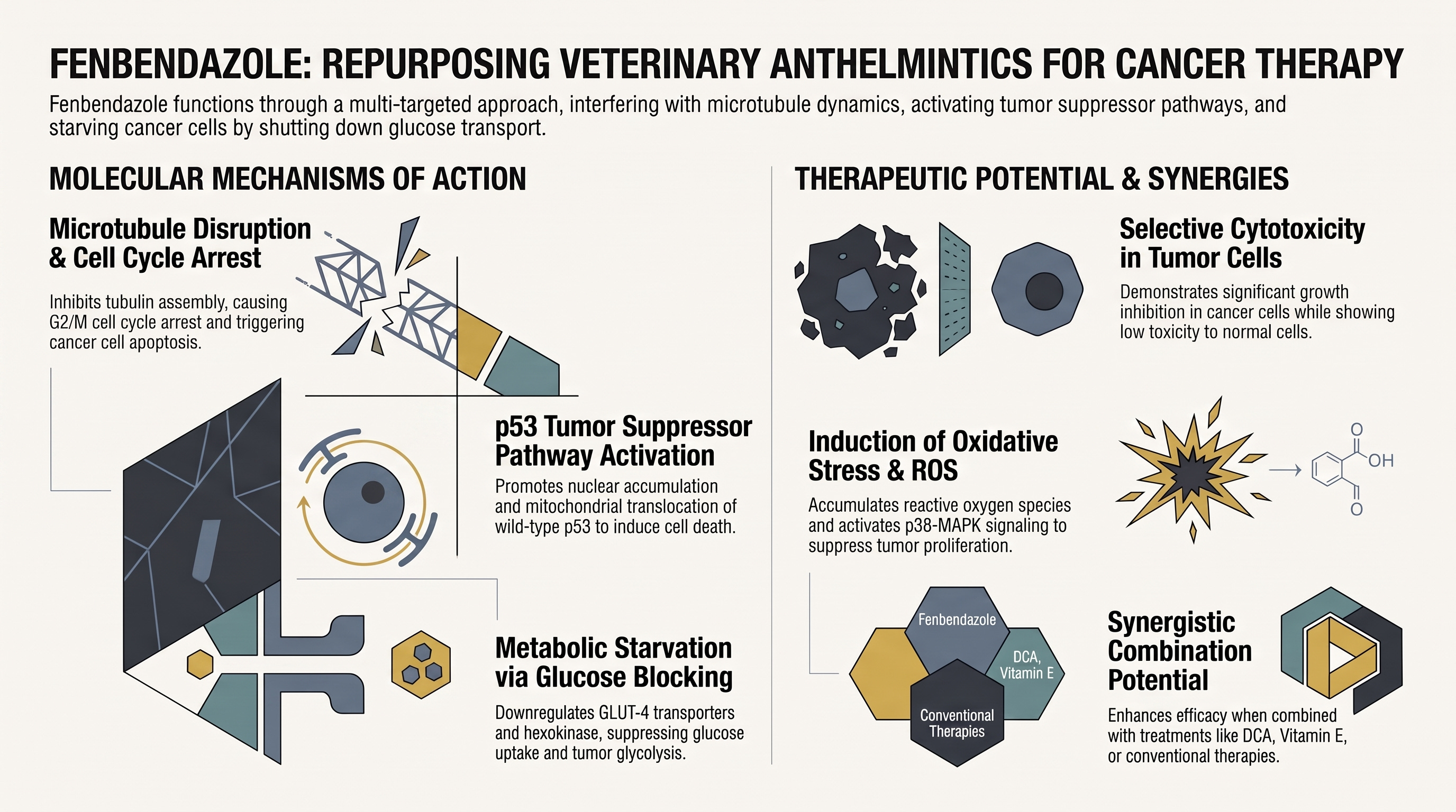
Glucose uptake inhibition is one of the primary mechanisms through which fenbendazole (FZ) starves cancer cells and sensitizes them to apoptosis.
Because malignant tumor cells rely heavily on accelerated aerobic glycolysis for ATP generation and cellular survival (the Warburg effect), interfering with their glucose supply causes metabolic stress that leads to cancer cell elimination.
Key Aspects of Fenbendazole-Induced Glucose Uptake Inhibition
1. Suppression of Glucose Import & Lactate Production
Reduced Glucose Uptake: Treatment with fenbendazole (1 µM) significantly inhibits the cellular uptake of the fluorescent glucose derivative 2-NBDG in human non-small cell lung carcinoma (NSCLC) cells (A549 and H460) within 4 hours.
Decreased Consumption & Lactate Release: FZ exposure leads to a dose-dependent reduction in overall glucose consumption and a corresponding drop in lactate secretion, indicating a severe disruption of glycolytic flux.
2. Downregulation of GLUT Transporters & Hexokinase II
GLUT-4 Downregulation: Fenbendazole significantly reduces the mRNA expression of the GLUT-4 glucose transporter, limiting the entry of glucose into tumor cells.
Hexokinase II (HKII) Impairment: FZ downregulates the gene expression and directly inhibits the enzymatic activity of Hexokinase II (HKII). HKII is the key rate-limiting enzyme that phosphorylates glucose to glucose-6-phosphate (G6P) for cellular retention and glycolytic processing.
Molecular Mimicry: In-silico docking and in-vitro enzymatic assays with cell extracts and purified enzyme demonstrate that fenbendazole stably binds to the glucose/G6P binding pocket of HKII, effectively acting as a molecular mimic that blocks normal enzyme function.
3. Interplay with p53 and Metabolic Regulators
p53-Mediated Repression: The inhibition of GLUT-4 and HKII expression following fenbendazole exposure is closely linked to p53 stabilization and microtubule alterations.
Induction of Metabolic Target Genes: While suppressing GLUT-4 and HKII, fenbendazole simultaneously induces p53-regulated metabolic and pro-apoptotic target genes, including TIGAR, SCO2, Glutaminase 2 (GLS2), and Proline Oxidase.
Enhancement of Succinate Dehydrogenase (SDH): FZ enhances the enzymatic activity of mitochondrial succinate dehydrogenase (SDH), a TCA cycle enzyme and tumor suppressor, altering metabolic flux away from harmful accumulation of succinate.
4. Synergistic Combination with Metabolic Inhibitors
Because fenbendazole suppresses glycolytic dependency, combining it with other metabolic agents produces strong synergistic anticancer effects (Combination Index < 1):
2-Deoxyglucose (2-DG): Co-treatment with 2-DG enhances glycolytic blockade and cell death.
Dichloroacetate (DCA): FZ synergizes strongly with DCA, a pyruvate dehydrogenase kinase inhibitor that forces cells away from glycolysis toward mitochondrial glucose oxidation.
In an in vivo study using female athymic nude mice (nu/nu) bearing human non-small cell lung carcinoma (A549) xenografts, oral administration of fenbendazole demonstrated significant antitumor efficacy.
The primary in vivo effects documented in the research include:
Marked Reduction in Tumor Size and Weight: Dosing mice orally with fenbendazole (1 mg per mouse in olive oil every second day for 12 days) led to a significant inhibition of tumor growth, resulting in markedly smaller tumor volumes and lower final tumor weights compared to untreated control mice.
Suppression of Tumor Angiogenesis & Vascularity: Fenbendazole significantly reduced tumor vascularity, as evidenced by a drop in intratumoral hemoglobin content. Immunohistochemical staining showed a sharp reduction in CD31-positive endothelial cells within the tumor xenografts, confirming anti-angiogenic activity.
Induction of Intratumoral Apoptosis: TUNEL staining of tumor tissue sections revealed a high density of apoptotic cells in the fenbendazole-treated group compared to controls.
In Vivo p53 Activation: Tumor sections from treated mice displayed a notable accumulation of p53-positive tumor cells, demonstrating that fenbendazole successfully triggers p53-mediated cell death pathways in living tumor tissue.
Overall, these findings confirm that the pleiotropic anti-cancer mechanisms observed in cell culture—such as microtubule destabilization, p53 activation, and anti-angiogenesis—translate into effective tumor growth suppression in vivo.
Fenbendazole Antitumor Efficacy
In an in vivo study using female athymic nude mice (nu/nu) bearing human non-small cell lung carcinoma (A549) xenografts, oral administration of fenbendazole demonstrated significant antitumor efficacy.
The primary in vivo effects documented in the research include:
Marked Reduction in Tumor Size and Weight: Dosing mice orally with fenbendazole (1 mg per mouse in olive oil every second day for 12 days) led to a significant inhibition of tumor growth, resulting in markedly smaller tumor volumes and lower final tumor weights compared to untreated control mice.
Suppression of Tumor Angiogenesis & Vascularity: Fenbendazole significantly reduced tumor vascularity, as evidenced by a drop in intratumoral hemoglobin content. Immunohistochemical staining showed a sharp reduction in CD31-positive endothelial cells within the tumor xenografts, confirming anti-angiogenic activity.
Induction of Intratumoral Apoptosis: TUNEL staining of tumor tissue sections revealed a high density of apoptotic cells in the fenbendazole-treated group compared to controls.
In Vivo p53 Activation: Tumor sections from treated mice displayed a notable accumulation of p53-positive tumor cells, demonstrating that fenbendazole successfully triggers p53-mediated cell death pathways in living tumor tissue.
Overall, these findings confirm that the pleiotropic anti-cancer mechanisms observed in cell culture—such as microtubule destabilization, p53 activation, and anti-angiogenesis—translate into effective tumor growth suppression in vivo.
Dogra, N., Kumar, A., & Mukhopadhyay, T. (2018). Fenbendazole acts as a moderate microtubule destabilizing agent and causes cancer cell death by modulating multiple cellular pathways. Scientific Reports, 8, Article 11926. https://doi.org/10.1038/s41598-018-30158-6