

How Dog Dewormer Fights Terminal Cancer (Audio)
1. Key Preclinical Mechanisms & Molecular Targets
1.1 Microtubule Dynamics and P-Glycoprotein (P-gp) Evasion
Fenbendazole (FBZ; methyl N-(6-phenylsulfanyl-1H-benzimidazol-2-yl) carbamate) is a broad-spectrum benzimidazole anthelmintic that exerts distinct anti-neoplastic activity through target interactions with mammalian tubulin dynamics while bypassing classical multidrug resistance mechanisms:
Colchicine-Binding Site Affinity: Competitive fluorescence-based binding assays demonstrate that FBZ binds to mammalian \alpha/\beta-tubulin heterodimers specifically at the colchicine-binding site, inhibiting subunit assembly into functional microtubules.
Moderate Microtubule Depolymerization: Unlike classical antimitotic agents such as colchicine and nocodazole, which cause severe, complete microtubule depolymerization into soluble tubulin subunits, FBZ shows moderate binding affinity and mild-to-moderate microtubule destabilization. Immunofluorescence analysis of human non-small cell lung carcinoma (NSCLC) A549 cells treated with 1 \mu\text{M} FBZ reveals a distorted, partially altered microtubule framework and loss of nuclear cage integrity without total cytoskeletal collapse. Quantitative fractionation assays demonstrate that the soluble-to-polymerized tubulin ratio (\text{Soluble}/\text{Polymerized} fold change) shifts modestly from 1.0 in control cells to \sim 1.5 in FBZ-treated cells, whereas nocodazole and colchicine induce severe shifts to \sim 2.0 and \sim 3.7, respectively (and paclitaxel reduces the ratio to \sim 0.75).
Maintenance of Acetylated Tubulin Levels: Western blot analyses using the 6-11B-1 antibody against acetylated \alpha-tubulin (Ac-\alpha-tubulin) show that while nocodazole, colchicine, and vincristine markedly deplete acetylated tubulin, FBZ maintains stable baseline levels of Ac-\alpha-tubulin. This preservation of tubulin acetylation maintains structural feedback signals essential for downstream stress transduction without precipitating immediate catastrophic collapse of non-mitotic cytoskeletal networks.
Bypass of P-Glycoprotein (P-gp/MDR-1) Efflux: Overexpression of P-glycoprotein (P-gp; encoded by MDR-1) represents a primary mechanism of acquired resistance to taxanes and vinca alkaloids. Experimental assays show that FBZ is neither a P-gp substrate nor an inhibitor. Co-administration of the P-gp inhibitor verapamil (10\,\mu\text{M}) does not alter FBZ-mediated inhibition of cancer cell proliferation. Furthermore, functional efflux assays monitoring intracellular accumulation of Rhodamine 123 (Rho123), a reference P-gp substrate, demonstrate no significant variance between control and FBZ-treated cells, confirming that FBZ bypasses P-gp-mediated drug clearance.
1.2 p53 Signaling Pathway, Translocation, and Apoptotic Cascades
The cytotoxic potency of FBZ is heavily governed by the functional status of the p53 tumor suppressor axis, operating through both transcriptional and direct mitochondrial pathways:
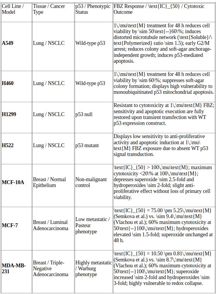
p53-Dependent Vulnerability: Human cancer cell lines with wild-type (WT) p53 (e.g., A549, H460) show greater sensitivity to FBZ-induced cytotoxicity than p53-null (H1299) or p53-mutant (H522) lines. Transient transfection of p53-null H1299 cells with a WT p53 expression vector fully restores sensitivity, resulting in robust apoptotic execution upon FBZ exposure.
Tubulin Acetylation and Nuclear/Mitochondrial Translocation: FBZ treatment induces dose-dependent nuclear accumulation of transcriptionally active WT p53. Mechanistically, FBZ preserves acetylated \alpha-tubulin levels, facilitating stress-mediated nuclear translocation of p53. Concurrently, FBZ impairs ubiquitin-proteasome system (UPS) degradation pathways, leading to accumulation of monoubiquitinated p53 that directly translocates to the outer mitochondrial membrane.
Mitochondrial Apoptotic Cascade: Mitochondrial accumulation of monoubiquitinated p53 disrupts membrane integrity, induces mitochondrial membrane depolarization (confirmed via JC-1 fluorometric ratio assays), releases cytochrome c, and triggers downstream cleavage of Poly (ADP-ribose) Polymerase (PARP). Simultaneously, nuclear p53 drives transcriptional activation of pro-apoptotic and metabolic target genes, including p21, GLS2 (glutaminase 2), Proline oxidase, TIGAR, and SCO2.
Early Mitotic Arrest and Caspase-Independent Death: FBZ accelerates and stabilizes cyclin B1/CDK1 complex formation (elevated at 8 hours post-treatment versus 16 hours in controls) and upregulates phosphorylated histone H3 at Ser10 [p-histone H3 (Ser10)], arresting cells in the G2/M phase. Subsequent cyclin B1 degradation at 32–48 hours coincides with a sub-G1 apoptotic peak (\sim 30\% at 32 hours) without polyploidy development. The broad-spectrum caspase inhibitor Z-VAD-FMK does not attenuate this mitotic death cascade, confirming a caspase-independent execution mechanism.
1.3 Metabolic Reprogramming and Glycolytic Pathways
FBZ systematically impairs malignant carbohydrate metabolism, targeting key steps of the Warburg effect:
Suppression of Glucose Transporters and Uptake: Treatment with 1\,\mu\text{M} FBZ significantly suppresses cellular glucose uptake in NSCLC models (measured by 2-NBDG fluorescent glucose uptake assays), decreases glucose oxidation rates, and reduces extracellular lactate accumulation.
Hexokinase II Active-Site Mimicry and Downregulation: FBZ downregulates mRNA expression of GLUT-4 and suppresses both mRNA expression and enzymatic activity of Hexokinase II (HK II). In silico molecular docking shows that FBZ structurally mimics glucose and glucose-6-phosphate (G6P), occupies the HK II catalytic active site pocket, and binds with high affinity to competitively block enzymatic phosphorylation.
Succinate Dehydrogenase (SDH) Tumor Suppressor Activation: In contrast to its known inhibition of parasite fumarate reductase (which converts fumarate to succinate in helminths), FBZ upregulates mitochondrial Succinate Dehydrogenase (SDH) activity in mammalian cells. SDH functions as a critical mitochondrial TCA cycle enzyme and tumor suppressor that catalyzes the oxidation of succinate to fumarate. By activating SDH, FBZ prevents the accumulation of succinate—an’oncometabolite’ that stabilizes HIF-1α and drives tumor progression—thereby restoring oxidative TCA flux and reversing aerobic glycolysis.
1.4 Selective Redox Modulation and Oxidative Stress Induction
FBZ displays differential, phenotype-dependent impact on intracellular reactive oxygen species (ROS) dynamics across malignant and non-malignant tissues:
Antioxidant/ROS Suppression in Normal Epithelium: In non-malignant breast epithelial cells (MCF-10A), 10\,\mu\text{M} FBZ exposure depresses basal oxidative stress. Dihydroethidium (DHE) fluorometric assays demonstrate a \sim 2.5-fold reduction in intracellular superoxide (\text{O}_2^{\bullet-}), while DCFH-DA assays demonstrate a \sim 2-fold reduction in hydroperoxides (\text{H}_2\text{O}_2) relative to untreated controls.
Pro-Oxidant ROS Augmentation in Malignant Tissues: In triple-negative breast cancer cells (MDA-MB-231), 10\,\mu\text{M} FBZ markedly amplifies cellular ROS, driving a \sim 2-fold surge in superoxide and a \sim 3-fold increase in hydroperoxides. In luminal adenocarcinoma cells (MCF-7), hydroperoxide levels rise \sim 1.5-fold while superoxide levels remain largely unchanged.
Warburg vs. Pasteur Phenotypic Vulnerability: This redox divergence stems from underlying metabolic wiring. Warburg-type cancer cells (glycolysis-dependent under normoxia and hypoxia, such as MDA-MB-231) lack metabolic plasticity, rendering them highly vulnerable to FBZ-induced ROS accumulation, ctDNA shedding, and rapid cytotoxic collapse. Conversely, Pasteur-type cells (oxidative phosphorylation-dependent under normoxia, such as MCF-7) maintain sufficient antioxidant buffer capacity to dampen ROS surges, resulting in moderate drug sensitivity.
2. In Vitro Cell Line Vulnerabilities and Cytotoxicity Profile
2.1 Comparative Cell Line Susceptibility Matrix
2.2 Subtype-Specific Vulnerabilities: Breast Cancer vs. Normal Epithelium
In vitro comparative profiles across breast epithelial models highlight significant lineage and metabolic dependencies:
Triple-Negative Breast Cancer Susceptibility (MDA-MB-231): Aggressive, triple-negative MDA-MB-231 cells exhibit high sensitivity (\text{IC}_{50} = 10.50 \pm 0.81\,\mu\text{M}). This susceptibility stems from their reliance on Warburg-type aerobic glycolysis, where FBZ-induced HK II inhibition and concurrent superoxide/hydroperoxide accumulation exceed lethal redox thresholds.
Luminal Adenocarcinoma Relative Resistance (MCF-7): Estrogen receptor-positive MCF-7 cells demonstrate lower baseline vulnerability (\text{IC}_{50} = 75.00 \pm 5.25\,\mu\text{M}). Pasteur-type metabolic flexibility mediates their relative resistance by relying on oxidative phosphorylation under normoxia and preventing a lethal intracellular ROS surge after FBZ exposure.
Nontransformed Epithelial Sparing (MCF-10A): Non-malignant MCF-10A cells exhibit safety margins (\text{IC}_{50} > 100\,\mu\text{M}), maintaining >80\% viability at 100\,\mu\text{M} drug concentration. FBZ acts as an antioxidant in non-malignant tissue, decreasing superoxide (\sim 2.5-fold) and hydroperoxide (\sim 2-fold) accumulation.
Resolution of Literature Discrepancies: Published literature contains contrasting \text{IC}_{50} metrics for MCF-7 cells (\sim 9.4\,\mu\text{M} reported by Vlachou et al. vs. 75.00 \pm 5.25\,\mu\text{M} reported by Semkova et al.). These divergent values reflect differences in primary cell stock origins, passaging history, baseline malignancy, divergent metabolic dependencies across sub-lines, and underlying drug-resistance mechanisms.
3. In Vivo Xenograft Outcomes and Chemo-Combination Strategies
3.1 In Vivo Efficacy in A549 Murine Xenografts
The in vivo antineoplastic activity of FBZ was established in female athymic nu/nu mice carrying established human NSCLC A549 xenografts (2\text{--}3\text{ mm} initial tumor diameter):
Dosing Protocol: Oral gavage of FBZ at 1\text{ mg/mouse} every second day for 12 days.
Tumor Weight and Volume Regression: FBZ treatment significantly inhibited macroscopic tumor volume growth and final excised tumor mass compared with vehicle controls.
Anti-Angiogenic Quantification: Spectrophotometric measurement of intratumoral hemoglobin content (\text{A}_{590\text{nm}}) demonstrated reduced intratumoral blood content. Immunohistochemical (IHC) analysis confirmed a marked reduction in CD31-positive microvessel endothelial cells within treated xenograft beds.
In Situ Apoptotic Induction & p53 Expression: Terminal deoxynucleotidyl transferase dUTP nick-end labeling (TUNEL) demonstrated a substantial increase in TUNEL-positive apoptotic cells in treated tumor tissue. Nuclear IHC staining confirmed elevated density of transcriptionally active p53-positive tumor cells, confirming in vivo execution of p53-mediated apoptosis.
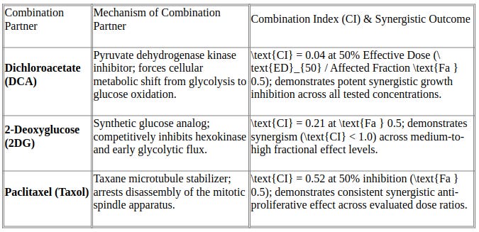
3.2 Quantitative Evaluation of Synergy with Metabolic and Chemotherapeutic Agents
4. Human Clinical Case Reports & Case Series Synthesis
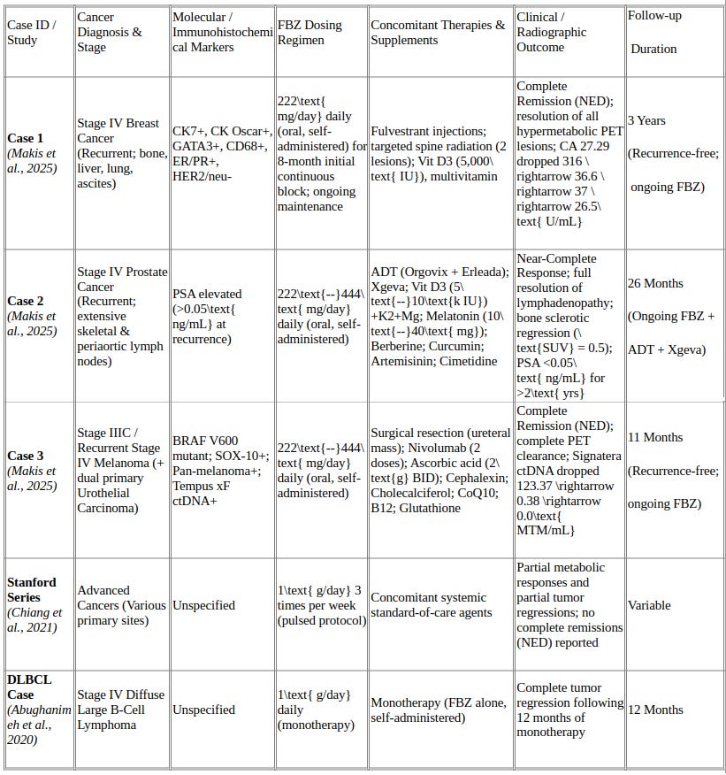
4.1 Comparative Matrix of Clinical Case Reports
4.2 Detailed Clinical Narrative and Biomarker Trajectories
Case 1: Stage IV Metastatic Breast Cancer
Patient Presentation & Baseline Diagnostics: An 83-year-old female originally diagnosed in 2009 with ER-positive breast cancer (treated via bilateral mastectomy, reconstruction, and aromatase inhibitors) presented with disease recurrence in October 2021. Liver fine needle aspiration (FNA) confirmed ER/PR-positive, HER2/neu-negative metastatic breast carcinoma (IHC: CK7+, CK Oscar+, GATA3+, CD68+). Malignant cells were present in ascitic fluid. Spine MRI revealed extensive osseous metastases across T10–L5, S1, S2, and iliac bones, including a 5.0 \times 2.9\text{ cm} lytic lesion in L4 extending into the spinal canal (\text{SUV}_{\text{max}} = 6.8) and a T12 lesion (\text{SUV}_{\text{max}} = 3.5). Baseline PET/CT (December 29, 2021) identified six hypermetabolic lung nodules (RUL index lesion 2.8 \times 1.5\text{ cm}, \text{SUV}_{\text{max}} = 8.4; RPL 1.8 \times 1.4\text{ cm}, \text{SUV}_{\text{max}} = 4.6; LUL 0.8\text{ cm}, \text{SUV}_{\text{max}} = 4.4) and hepatic lesions (2.9 \times 1.7\text{ cm}, \text{SUV}_{\text{max}} = 5.6).
Therapeutic Timeline & Biomarker Clearance: The patient declined conventional multi-agent chemotherapy and radiation and entered hospice care. On November 22, 2021, she initiated self-administered oral FBZ at 222\text{ mg/day}. In December 2021, she started fulvestrant injections to restrict estrogen-driven metabolic supply, complementing FBZ’s inhibition of GLUT-4/HK II glucose entry. In January 2022, targeted radiation was delivered to two painful spinal lesions. The patient maintained an 8-month continuous block of FBZ (222\text{ mg/day}). CA 27.29 tumor markers exhibited a precipitous decline: 316\text{ U/mL} (Nov 2021) \rightarrow 36.6\text{ U/mL} (July 2022) \rightarrow 37\text{ U/mL} (July 2022) \rightarrow 26.5\text{ U/mL} (Feb 2023; normal range). Serial PET/CT imaging in April and June 2022 confirmed complete resolution of hypermetabolic activity across all pulmonary, hepatic, and osseous lesions, establishing Complete Remission (”No Evidence of Disease”). All active interventions were discontinued in June 2022; the patient remains recurrence-free nearly 3 years post-remission on maintenance FBZ.
Case 2: Stage IV Recurrent Prostate Cancer
Patient Presentation & Baseline Diagnostics: A 75-year-old male diagnosed in 2011 with prostate cancer (surgically treated) maintained undetectable PSA levels for 18 months post-surgery before a rising PSA heralded metastatic recurrence in December 2021. Abdominal/pelvic CT (December 16, 2021) identified left periaortic lymphadenopathy (0.8\text{ cm}). Bone scintigraphy confirmed widespread osteoblastic lesions involving the spine, pelvic ring, and right humeral head.
Therapeutic Timeline & Radiographic Resolution: In December 2021, the patient initiated double-androgen deprivation therapy (ADT; Orgovix and Erleada), denosumab (Xgeva), and daily oral FBZ (222\text{--}444\text{ mg/day}), alongside targeted supplements (vitamin D3 5,000\text{--}10,000\text{ IU/day} + K2 + Mg, melatonin 10\text{--}40\text{ mg}, berberine, curcumin, artemisinin, cimetidine). Follow-up CT scans at 12 months (December 2022) and 24 months (January 2024) demonstrated complete regression of periaortic lymph nodes and progressive sclerotic healing of bone lesions without new foci. A whole-body PSMA-PET/CT scan (April 2024) confirmed non-avid sclerotic bone lesions, absence of nodal radiopharmaceutical uptake, and a background renal cortical cyst (\text{SUV} = 0.5). Serum PSA remained suppressed below detection limits (<0.05\text{ ng/mL}) for >2\text{ years}. The patient achieved a Sustained Near-Complete Response over 26 months of ongoing FBZ and ADT/Xgeva therapy.
Case 3: Stage IIIC / Recurrent Stage IV Melanoma
Patient Presentation & Baseline Diagnostics: A 63-year-old male diagnosed in July 2020 with BRAF V600-mutated Stage IIIC melanoma received adjuvant dabrafenib and trametinib, which was discontinued early in May 2021 due to a reduced left ventricular ejection fraction. Remission persisted until December 12, 2023, when a biopsy of a new 1.6\text{ mm} ulcerated abdominal lesion confirmed recurrent melanoma (SOX-10+, pan-melanoma+). Baseline PET/CT showed hypermetabolic retroperitoneal/peritoneal nodules, stomach, small bowel, gluteus medius, quadratus femoris, and L5 osseous foci. Biopsy of an incidental distal ureteral mass revealed a distinct secondary primary urothelial carcinoma. Tempus xF liquid biopsy detected circulating BRAF V600 mutant ctDNA.
Therapeutic Timeline & ctDNA Kinetics: Systemic immunotherapy was delayed to allow surgical resolution of ureteral obstruction. During this treatment-free window in mid-December 2023, the patient initiated self-administered FBZ (222\text{--}444\text{ mg/day}). He subsequently underwent ureteral surgery and received two doses of nivolumab. Serial ctDNA monitoring (Signatera) revealed rapid molecular clearance: 123.37\text{ MTM/mL} (Nov 29, 2023; pre-FBZ) \rightarrow 0.38\text{ MTM/mL} (Jan 17, 2024; <7 weeks on FBZ) \rightarrow 0.0\text{ MTM/mL} (Feb 21, 2024). This dramatic clearance reflects the vulnerability of highly glycolytic, Warburg-phenotype melanoma cells to FBZ-induced ROS surges and metabolic blockade. Follow-up PET/CT imaging in February 2024 confirmed Complete Remission (”No Evidence of Disease”). The patient remains recurrence-free over 11 months post-remission on daily maintenance FBZ.
5. Toxicological Profile, Comparative Limitations, and Regulatory Imperatives
5.1 Toxicity, Safety Margins, and Hepatic Function
Preclinical models establish high therapeutic safety margins for FBZ in non-malignant tissues (MCF-10A \text{IC}_{50} > 100\,\mu\text{M}; primary rat lung epithelial viability >70\text{--}90\%). In human clinical case reports, daily doses of 222\text{ mg/day} to 444\text{ mg/day} were generally well tolerated without subjective systemic toxicities.
However, clinical biomarker tracking in Case 1 identified a transient elevation of serum alanine aminotransferase (ALT) and aspartate aminotransferase (AST) in July 2022. Distinguishing the precise etiology of this transient transaminitis presents diagnostic challenges, with potential contributing factors including:
Direct FBZ-mediated hepatic metabolism and microsomal enzyme interaction.
Fulvestrant-induced hepatic inflammation.
Competitive cytochrome P450 drug-drug interactions (e.g., CYP2C19/CYP2J2 substrate pathways) between FBZ and fulvestrant.
Transaminase elevations normalized within weeks without permanent hepatic damage or treatment cessation.
Dosing schedules vary across published human reports. The continuous daily low-dose regimen (222\text{--}444\text{ mg/day}) evaluated by Makis et al. contrasts with the pulsed protocol (1\text{ g/day} 3 times per week) reported in the Stanford case series by Chiang et al., and the continuous high-dose monotherapy (1\text{ g/day} daily) documented by Abughanimeh et al. Controlled clinical settings have not characterized the comparative human pharmacokinetics, oral bioavailability, tissue distribution, and long-term hepatic safety profiles of these regimens.
5.2 Methodological Limitations and Therapeutic Confounders
The existing human clinical evidence evaluating FBZ in oncology contains severe methodological limitations:
Small Sample Size and Observational Nature: Published human clinical data are limited to isolated case reports and small retrospective series (n=3 in Makis et al.; n=1 in Abughanimeh et al.). These observational cohorts lack statistical power, comparative control arms, and generalizability to broader patient populations.
Confounding Concomitant Therapies: In virtually all clinical cases, patients received concurrent modalities, including anti-estrogen therapy (fulvestrant), androgen deprivation therapy (Orgovix, Erleada), bisphosphonates/RANKL inhibitors (Xgeva), targeted palliative radiation, surgical resections, immune checkpoint inhibitors (nivolumab), and aggressive multi-agent nutraceutical supplementation. These co-interventions make it impossible to isolate FBZ's individual antineoplastic contribution or prove direct causality.
Self-Selection and Reporting Bias: Published reports involve patients who independently initiated veterinary-grade or off-label formulations. Retrospective literature selection is highly susceptible to publication bias, where favorable clinical responses are preferentially reported while non-responses, rapid disease progressions, or adverse toxicities remain undocumented.
Lack of Methodological Rigor and Regulated Controls: Clinical data are observational and lack the controlled parameters of prospective clinical trials, such as standardized protocol dosing, serial pharmacokinetic/pharmacodynamic blood sampling, structured phase-specific toxicity grading, and objective radiological verification using Response Evaluation Criteria in Solid Tumors (RECIST 1.1).
5.3 Risks of Patient-Led Repurposing and Future Regulatory Directions
The increasing trend of patient-led self-medication using veterinary-grade or unregulated off-label compounds introduces significant clinical risks. Sourcing compounds outside formal pharmaceutical supply chains exposes patients to unverified active pharmaceutical ingredient (API) purity, variable excipient profiles, unstandardized dosing, unmonitored drug-drug interactions, and potential unmanaged hepatic or systemic toxicities.
To transition from anecdotal observation to evidence-based oncology, formal, phase-structured human clinical trials are urgently required. Well-designed prospective trials are essential to establish definitive safety and tolerability limits, define optimal clinical dosing schedules and human pharmacokinetics, and determine whether FBZ possesses true single-agent or combination efficacy in human oncology.
References
Abughanimeh, O., Evans, T., & Kallam, A. (2020). Fenbendazole as a treatment for diffuse large B-cell lymphoma. Annals of Hematology & Oncology, 7(2), Article 1284.
Dogra, N., Kumar, A., & Mukhopadhyay, T. (2018). Fenbendazole acts as a moderate microtubule destabilizing agent and causes cancer cell death by modulating multiple cellular pathways. Scientific Reports, 8(1), Article 11926. https://doi.org/10.1038/s41598-018-30158-6
Khattab, M., & Al-Karmalawy, A. A. (2021). Revisiting activity of some nocodazole analogues as a potential anticancer drugs using molecular docking and DFT calculations. Frontiers in Chemistry, 9, Article 628398.
Makis, W., Baghli, I., & Martinez, P. (2025). Fenbendazole as an anticancer agent? A case series of self-administration in three patients. Case Reports in Oncology, 18(1), 864–875. https://doi.org/10.1159/000546362
Park, D., Lee, J.-H., & Yoon, S.-P. (2022). Anti-cancer effects of fenbendazole on 5-fluorouracil-resistant colorectal cancer cells. Korean Journal of Physiology & Pharmacology, 26(5), 377–387. https://doi.org/10.4196/kjpp.2022.26.5.377
Semkova, S., Nikolova, B., Tsoneva, I., Antov, G., Ivanova, D., Angelov, A., Zhelev, Z., & Bakalova, R. (2023). Redox-mediated anticancer activity of anti-parasitic drug fenbendazole in triple-negative breast cancer cells. Anticancer Research, 43(3), 1207–1212.